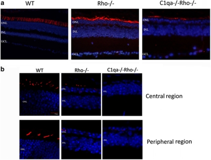
Figure 2.
Comparative retinal histology in WT, Rho−/−, and Rho−/−C1qa−/− mice. (a) The presence and pattern of cone photoreceptors were analyzed in WT, Rho−/−, and C1qa−/−Rho−/− mice. Although there was positive peanut agglutinin staining in Rho−/− and C1qa−/−Rho−/− mice at 12 weeks of age, the pattern and distribution of staining appeared radically different in C1qa−/−Rho−/− mice when compared with Rho−/− mice. (b) Retinal cryosections from 12-week-old mice were stained with an antibody specific for blue-sensitive opsin. Strong immunoreactivity was observed in the WT sections, staining blue cone photoreceptors in the central and peripheral aspects of the retina, and clearly showing the distribution of cone photoreceptors in the mouse retina (red: blue-sensitive opsin; blue: DAPI-nuclei). Although not as widespread, positive immunoreactivity for blue-sensitive opsin was also evident in Rho−/− mice. However, in C1qa−/−Rho−/− mice, strong immunoreactivity in cryosections for blue-sensitive opsin was not evident.

