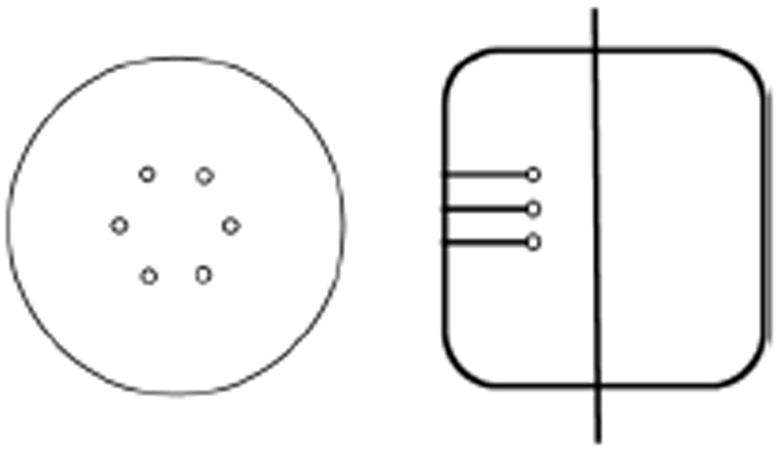
Fig. 1.

Transverse and axial views of the 35-cm diameter by 30-cm long lesion phantom. The spheres are arranged at a fixed radial position and placed in a plane at ¼ the axial length of the scanner (vertical line in axial view shows schematically the central plane of the scanner).
