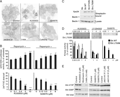
FIGURE 3.
KU55933 and Gö6976 inhibit Vps34. A, confocal microscopy images of GFP-2XFYVE expressing MCF7 cells either untreated (Control) or treated for 2 h with 2 μm KU55933, 8 μm Gö6976, 5 μm Jak3 inhibitor VI, 10 mm 3-MA, or 10 mm caffeine. The images were converted to grayscale and inverted using Photoshop. B, comparison of the effects of KU55933 (left) and Gö6976 (right) on rapamycin-induced autophagy and on eGFP-2XFYVE-relocalization. (B, top panels) MCF7 cells expressing RLucLC3wt or RLuc-LC3G120A were left untreated or treated with 100 nm rapamycin alone or in combination with indicated concentrations of KU55933 or Gö6976 for 6 h. The data represent average ± S.D. of at least three independent experiments. (B, bottom panels) MCF7- eGFP-2XFYVE cells were treated with indicated concentrations of KU55933 or Gö6976 for 2 h and the number of eGFP-positive dots/cell were calculated in confocal images. *, p value <0.05; **, p value <0.01; and ***, p value <0.001 as compared with cells treated with rapamycin alone (B, top) or control (B, bottom). C, immunoblotting with antibodies against Vps34 and Beclin 1 of streptactin-pulldowns of MCF7 cells transiently transfected with pEXPR-IBA105 (Str.), pStrep-VPS34wt (Str.WT), pStrep-VPS34D743N (Str. D743N), or pStrep-VPS34L750M (Str. L750M). The bottom row shows immunoblotting of the corresponding lysates with antibody against Beclin 1. D, dot blotting of PtdIns(3)P produced in vitro by strep-tagged Vps34WT (Str.WT) or strep-tagged Vps34L750M (Str.L750M). Kinase reactions were performed in the absence of inhibitors (Control) or with either KU55933, LY-294002, or Gö6976 at the indicated concentrations. The bar graph shows the Quantification of PtdIns(3)P dot-blots from Vps34 in vitro kinase reactions analyzing either Strep-tagged Vps34wt or Strep-tagged Vps34L750M. Kinase reactions were performed in the absence of inhibitors (Control) or with KU55933 (n = 3), LY-294002 (Vps34wt) (n = 3) LY-294002 (Vps34L750M) (n = 2), or Gö6976 (n = 2) as indicated. E, immunoblotting with antibodies against Akt and phospho-Akt in total extracts from MCF7 cells incubated with the indicated concentrations of inhibitors for 6 h.

