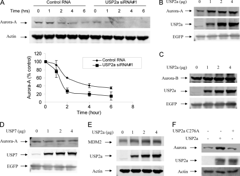
FIGURE 2.
USP2a regulates the protein levels of Aurora-A. A, MIA PaCa-2 cells were transfected with 30 nm USP2a siRNA for 72 h. The cells were then treated with 50 μm cycloheximide for the indicated time. The cells were harvested, and Western blot analysis was carried out for Aurora-A and actin. The lower panel shows the quantification of the levels of Aurora-A protein. The intensity of protein band was determined using a Bio-Rad GS-800 calibrated densitometer (Bio-Rad) and normalized according to actin. Data are the average of two independent experiments. Error bars represent S.D. B, H1299 cells were transfected with 0.5 μg of pcDNA3-EGFP and 1 μg of pc-DNA3-Aurora-A in the presence of 0, 1, 2, or 4 μg of pCMV-xl6-USP2a for 24 h. The cells were treated with 20 μm cycloheximide for 4 h. Cells were harvested, and the cell lysates were prepared and subjected to Western blot analysis for Aurora-A, USP2a, and EGFP. C, experiments similar to those in B were carried out except 1 μg of pc-DNA3-Myc-Aurora-B was used. D, experiments similar to those in B were carried out except pc-DNA3-Myc-USP7 was used. E, experiments similar to those in B were carried out except 1 μg of pc-DNA3-Myc-MDM2 was used. F, H1299 cells were transfected with 1 μg of pc-DNA3.1 (−) Myc-Aurora-A and 4 μg of pCMV-xl6-USP2a or 4 μg of pcDNA3-USP2a-C276A for 24 h. Cells were harvested, and lysates were prepared and subjected to Western blot analysis for Aurora-A, USP2a, and actin.

