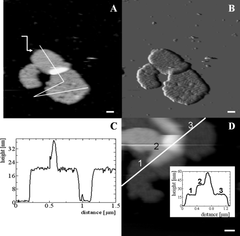
FIGURE 2.
AFM images of grana membranes adhered to mica and stored at 4 °C prior to rinsing and imaging. A, height image. The white line indicates the position of the height profile presented in C. The arrow points to an empty piece of membrane. B, error image of A. C, height profile as indicated in A. The zero level is taken at the mica surface. D, height image and profile (inset) of three pieces of membrane on top of each other. Scale bars = 100 nm.

