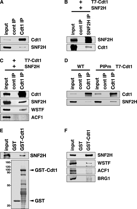
FIGURE 1.
Cdt1 interacts with SNF2H. A, nuclear extracts were prepared from HEK293T cells and immunoprecipitated with anti-Cdt1 antibody (Cdt1 IP) or control rabbit IgG (cont IP). Immunoprecipitates were subjected to immunoblotting with the indicated antibodies. Three percent of the input was also loaded. B, HEK293T cells were transfected with T7-Cdt1 and SNF2H expression vectors, and nuclear extracts were prepared. After immunoprecipitation with anti-SNF2H antibody, the precipitates were blotted with the indicated antibodies. One percent of the input for SNF2H or 0.3% of the input for Cdt1 was loaded. C, HEK293T cells were transfected with T7-Cdt1 and SNF2H expression vectors, and nuclear extracts were prepared. After immunoprecipitation with anti-Cdt1 antibody, the precipitates were immunoblotted with the indicated antibodies. One percent of the input was loaded. D, HEK293T cells were transfected with the indicated expression vectors, and nuclear extracts were prepared. After immunoprecipitation with anti-Cdt1 antibody or control rabbit IgG, the precipitates were immunoblotted with the indicated antibodies. Three percent of the input was loaded. E, GST-Cdt1 or GST was incubated with SNF2H protein synthesized by in vitro transcription-translation with rabbit reticulocyte lysate. GST-Cdt1 and the associated SNF2H protein were then collected on glutathione beads and subjected either to immunoblotting with anti-SNF2H antibody (upper panel) or to Coomassie Brilliant Blue staining (lower panel). Twenty percent of the input sample was analyzed. F, GST-Cdt1 or GST was incubated with HeLa nuclear extracts, and bound proteins were analyzed by immunoblotting with the indicated antibodies. Fifteen percent of the input was loaded.

