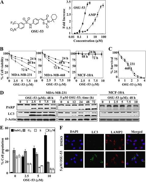
FIGURE 1.
OSU-53 activates AMPK, inhibits breast cancer cell proliferation, and induces apoptosis and autophagy. A, left panel, structure of OSU-53. Right panel, effect of OSU-53 on the kinase activity of recombinant AMPK. Kinase activity was determined in the presence of OSU-53 or AMP at the indicated concentrations by measuring 32P-phosphorylation of the SAMS peptide substrate as described under “Experimental Procedures.” Points, mean; bars, S.D. (n = 3). B, effects of OSU-53 on the viability of MDA-MB-231 and MDA-MB-468 breast cancer cells and MCF-10A breast epithelial cells. Cells were treated with OSU-53 at the indicated concentrations in 5% FBS-supplemented medium for 24, 48, and 72 h, and cell viability was assessed by MTT assays. Points, mean; bars, S.D. (n = 6). C, effects of OSU-53 on clonogenic survival of breast cancer cell lines (231, MDA-MB-231; 468, MDA-MB-468). Cells were treated with OSU-53 at the indicated concentrations for a total of 10–12 days, then fixed, stained, and counted as described under “Experimental Procedures.” Points, mean; bars, S.D. (n = 3). D, Western blot analysis of the concentration- and/or time-dependent effects of OSU-53 on PARP cleavage, a marker of apoptosis, and LC3-II conversion, a marker of autophagy, in MDA-MB-231 and MCF-10A cells. E, dose-dependent effect of OSU-53 on cell cycle distribution in MDA-MB-231 cells after 48 h of treatment. Data are presented as means ± S.D. of three independent experiments. F, effect of OSU-53 on autophagosome formation in MDA-MB-231 cells. Cells were treated with 5 μm OSU-53 for 48 h and then stained for LC3 and LAMP2 as described under “Experimental Procedures.” Colocalization of LC3 with LAMP2 was visualized by confocal microscopy. Green, LC3 (Alexa Fluor 488); red, LAMP2 (Alexa Fluor 555); blue, nuclei (DAPI). Magnification, ×40.

