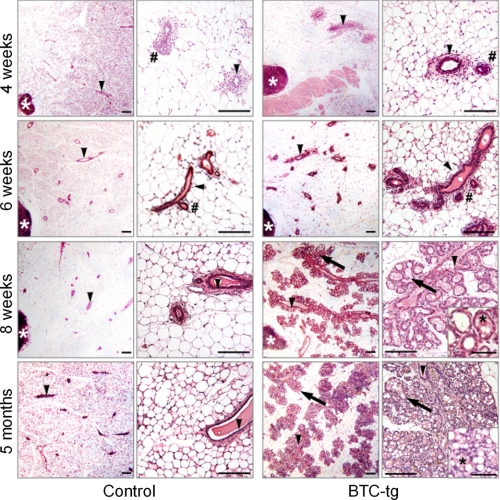
FIGURE 2.
Histology of mammary gland development in non-transgenic control mice (left column) and BTC-tg mice (right column) at 4, 6, and 8 weeks, and at 5 months of age. Per genotype and age, a survey view (left, bar = 200 μm) and a higher magnification (right, bar = 100 μm) are shown. The histology of alveolar epithelial cells of BTC-tg mice is shown in inset pictures for BTC-tg mice at 8 weeks and 5 months of age (bar = 50 μm). Lymph nodes (white asterisks), lactiferous ducts (arrowheads), end/side buds (#), alveolar lobules and acini (arrows), and milk-like secretion (black asterisks) are indicated. Paraffin sections of the fourth abdominal mammary gland complexes, H&E staining.

