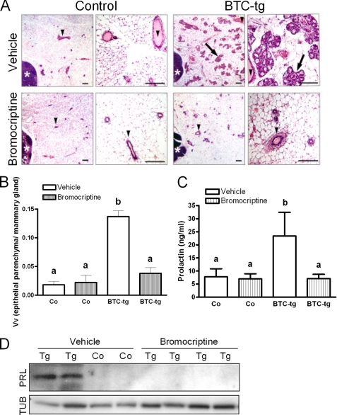
FIGURE 5.
A, histology of the fourth abdominal mammary gland complex of control females and BTC-tg females treated with vehicle (upper panels) or bromocriptine (lower panels) for 11 weeks. Per genotype, a survey view (left, bar = 200 μm) and a higher magnification (right, bar = 100 μm) are shown. Lymph nodes (white asterisks), lactiferous ducts (arrowheads), alveolar lobules (arrows). Paraffin sections, H&E staining. B, volume densities of mammary epithelial parenchyma (lactiferous ducts, alveolar buds, lobules, and acini) in the mammary gland (VV(mammary epithelial parenchyma/mammary gland)) of the same animals (n = 3–5/group). C, while bromocriptine treatment did not alter serum prolactin levels in control mice, it significantly reduced the serum levels of the hormone in BTC-tg mice (same animals as above). D, Western blot analysis showing that bromocriptine treatment abolished the occurrence of prolactin (PRL) in the mammary glands of BTC-tg females. TUB: tubulin. Data in B and C are means ± S.D. Significant differences (B: p < 0.001, C: p < 0.01) between the respective groups are indicated by different superscripts (a, b).

