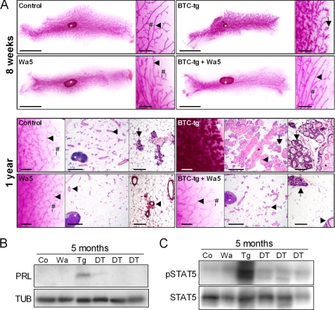
FIGURE 6.
A, mammary gland morphology (4th abdominal mammary gland complexes) of control, BTC-tg, Wa5 and BTC/Wa5 double-transgenic mice. Top panel, mammary gland whole mount preparations of 8 weeks old mice. Per genotype, a survey view (left, bar = 1 cm) and a higher magnification (right, bar = 1 mm) are shown. Bottom panel: whole mount preparations and histology (paraffin sections, H&E staining) of mammary glands of 1-year-old mice. Per genotype, a higher magnification of a whole mount preparation (right, bar = 1 mm), a survey view of a histological section (middle, bar = 1 mm) and a higher magnification of a histological section (right, bar = 100 μm) are shown. Lymph nodes (white asterisks), lactiferous ducts (arrowheads), side buds (#), alveolar buds, lobules, and acini (arrows), and milk-like secretion (black asterisks) are indicated. Note the reduction of hyperplastic growth of alveolar acini in BTC/Wa5-tg mice as compared with BTC-tg mice. B, Western blot showing that the presence of prolactin in the mammary gland, visible in BTC-tg females, disappears after crossing into the EGFR dominant-negative background Wa5 (DT, double transgenic mice). C, Western blot showing the disappearance of phosphorylated STAT5 in the mammary gland of BTC-tg females after crossing into the EGFR dominant-negative background Wa5 (DT, double transgenic mice).

