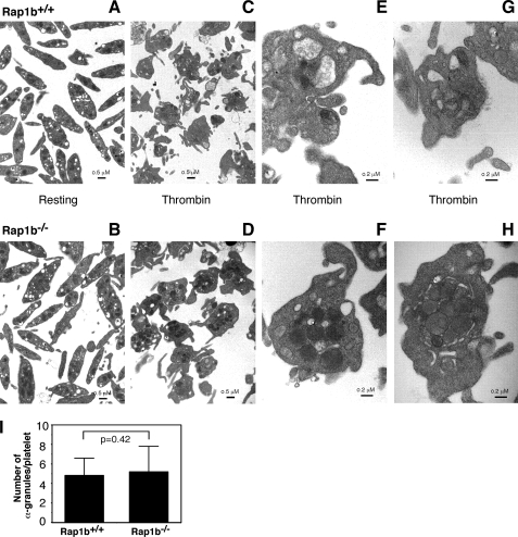
FIGURE 3.
Platelet electron microscopy analysis. A, washed platelets from Rap1b+/+ (A, C, E, and G) and Rap1b−/− (B, D, F, and H) mice were either maintained in resting state or incubated with 0.1 unit/ml thrombin for 5 min at 22 °C. The samples were fixed and analyzed by transmission electron microscopy. A and B represent platelets under resting conditions, and C–H represent platelets stimulated with thrombin. Scale bars represent 0.5 μm for A–D, and 0.2 μm for E–H. I, numbers of α granules in all platelets from five randomly chosen fields of Rap1b−/− (5.20 ± 2.61/platelets, n = 75) and Rap1b+/+ (4.82 ± 1.77/platelets, n = 84) resting samples were counted. Statistical significance was determined using Student t test.

