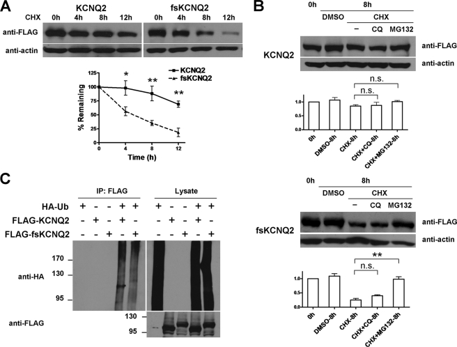
FIGURE 4.
Accelerated degradation of fsKCNQ2 mediated by the ubiquitin-independent proteasome pathway. A, shown is a CHX treatment assay of KCNQ2 and fsKCNQ2 in Cos-7 cells. Cells were transfected with plasmids FLAG-KCNQ2 or FLAG-fsKCNQ2 and treated with protein synthesis inhibitor CHX (75 μg/ml) for the indicated time period (0, 4, 8, or 12 h). After degradation, the remaining proteins were separated by SDS-PAGE and analyzed by Western blotting using anti-FLAG or anti-actin antibody. Data are expressed as the mean ± S.E. (n = 4; *, p < 0.05; **, p < 0.01). The half-life of KCNQ2 was more than 12 h, and the half-life of fsKCNQ2 was about 4 h. B, shown is a Western blotting analysis of KCNQ2 and fsKCNQ2 in Cos-7 cells treated with CHX (75 μg/ml) and chloroquine (CQ, 50 μm) or MG132 (15 μg/ml). Samples were loaded in lanes from the left in the following order: control without any treatment, DMSO treatment for 8 h, CHX for 8 h, CHX with chloroquine for 8 h, CHX with MG132 for 8 h. Data were normalized to that of control without any treatment and are expressed as the mean ± S.E. (n = 3, **, p < 0.01, n.s., not significant, p > 0.05). C, Cos-7 cells were transfected with combinations of FLAG-KCNQ2 or FLAG-fsKCNQ2 and HA-ubiquitin plasmids as indicated with a plus sign and treated with MG132 for 12 h. Cell lysates were subjected to immunoprecipitation (IP) by anti-FLAG antibody before Western blotting with anti-HA antibody. As a loading control, lysates were directly Western-blotted with anti-HA or anti-FLAG antibody.

