FIGURE 4.
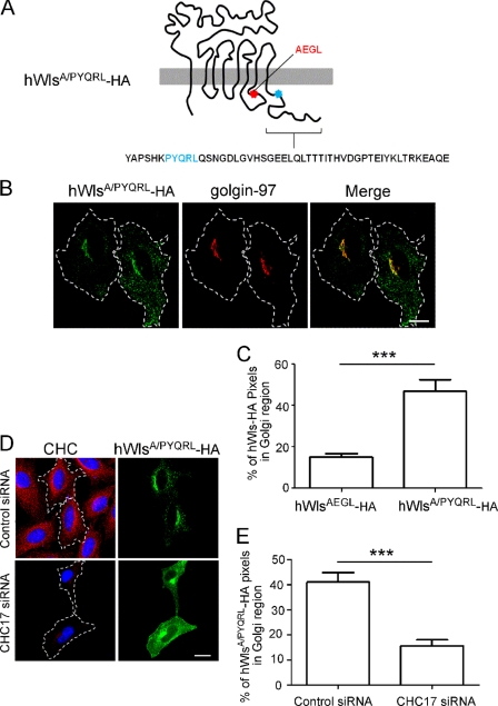
The hWlsAEGL-HA mutant is rescued with a functional YXXφ motif inserted into the cytoplasmic tail. A, hWlsA/PYQRL-HA construct is diagrammed. B, HeLa cells were transfected with hWlsA/PYQRL-HA for 24 h, and monolayers were fixed, permeabilized, and stained with monoclonal anti-HA antibodies (green) and monoclonal anti-golgin-97 antibodies (red). D, HeLa cells were transfected with control siRNA or CHC17 siRNA for 48 h followed by transfection with hWlsA/PYQRL-HA for a further 24 h. Cells were fixed and permeabilized and stained with monoclonal anti-HA antibodies (green) and monoclonal antibodies to clathrin heavy chain (CHC) (red). Nuclei were stained with DAPI. Scale bars represent 10 μm. C and E, hWlsAEGL-HA and hWlsA/PYQRL-HA were quantified at the Golgi (C) and hWlsA/PYQRL-HA at the Golgi of siRNA-treated cells (E). Shown are the proportion of HA pixels within the Golgi region relative to the total number of HA pixels/cell, expressed as the mean ± S.E. (error bars) (n = 20 for each dataset). ***, p < 0.001.

