FIGURE 5.
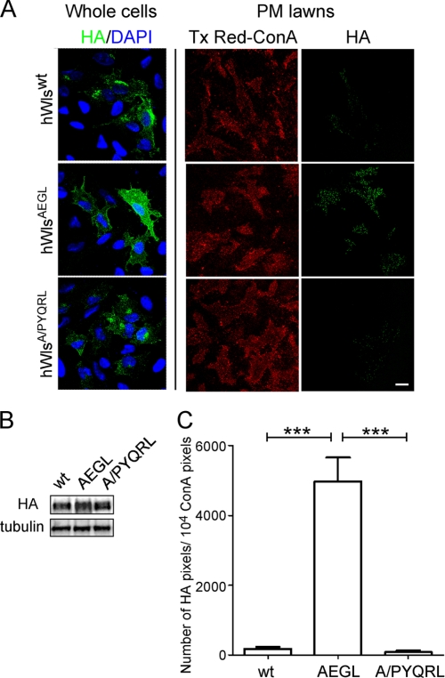
The hWlsA/PYQRL mutant rescues the AEGL internalization defect. A, HeLa cells were seeded on poly-l-lysine coverslips and transfected with hWlswt-HA, hWlsAEGL-HA, or hWlsA/PYQRL-HA for 24 h. Monolayers were either directly fixed and permeabilized (whole cells) and stained with monoclonal HA antibodies (green) and DAPI (blue) or treated with hypotonic buffer (PM lawns) followed by fixation and stained with Tx Red-ConA (red) and monoclonal HA antibodies (green). Scale bar represents 10 μm. B, immunblotting of transfected cell populations from A. Transfected monolayers were lysed in SDS-PAGE reducing buffer and cell extracts subjected to SDS-PAGE on a 4–12% gradient polyacrylamide gel. Proteins were transferred to a PVDF membrane and probed with rabbit anti-HA antibodies using a chemiluminescence detection system. The membrane was then stripped and reprobed with mouse anti-α-tubulin antibodies. C, the number of HA pixels in transfected, sonicated cells was quantified. Data are expressed as the mean number of HA pixels/104 Tx Red-ConA pixels, ± S.E. (error bars) (n = 20 for each dataset) and normalized for densitometry values from the HA Western blot. ***, p < 0.001.

