
FIGURE 2.
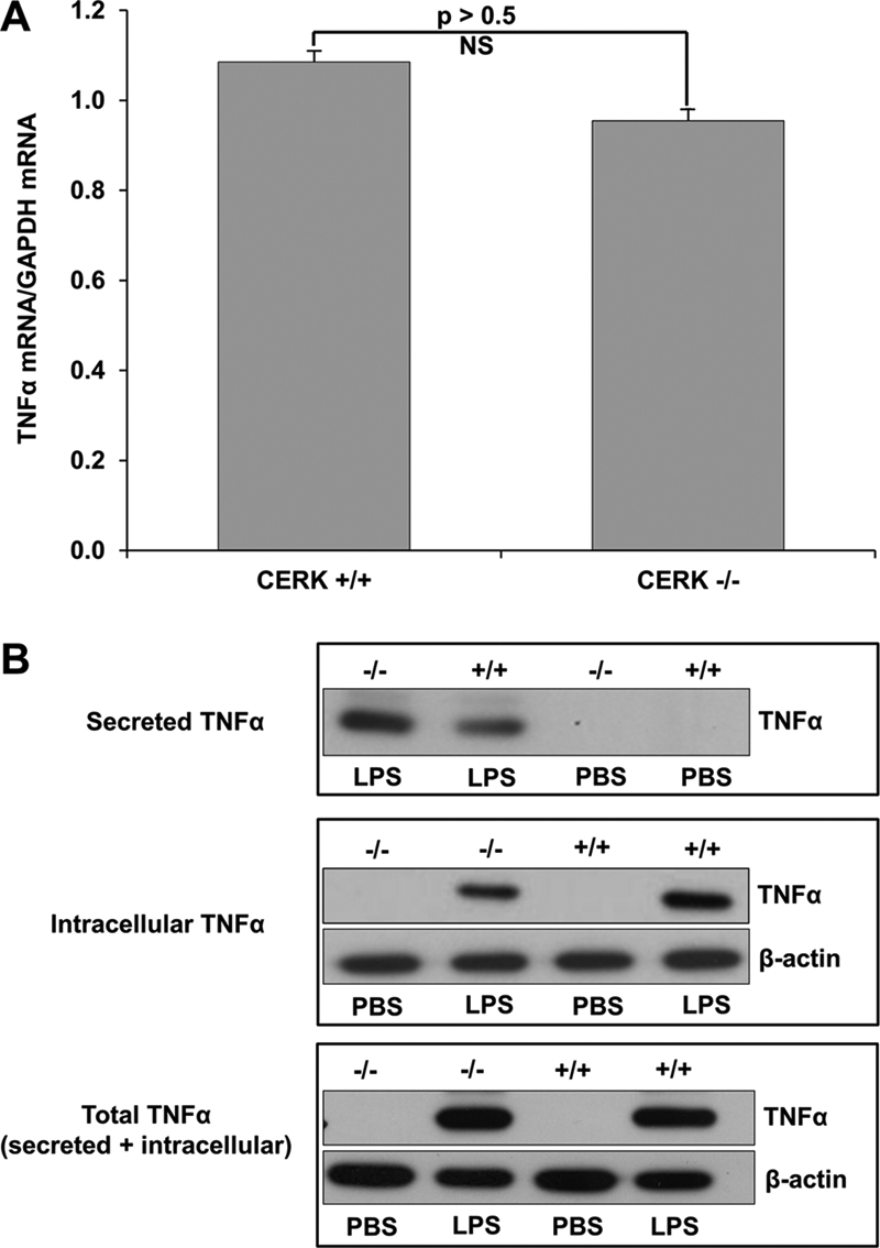
TNFα secretion but not expression is significantly different in CERK null macrophages compared with wild type. Primary monocytes isolated from the bone marrow of CERK null mice and their wild-type littermates were differentiated into BMDMs as described under “Experimental Procedures.” The BMDMs (5 × 10⁵ cells), thus obtained, were seeded overnight in 35-mm plates. Cells were then treated with LPS (0.5 ng/ml) or PBS sham, and total RNA and protein were harvested 4 h post-treatment. A, TNFα and GAPDH mRNA levels were quantified via quantitative PCR with TNFα mRNA levels normalized to GAPDH expression. Data presented in this figure are the means of three separate experiments ± S.E. (n = 3). B, protein extracts from cells (10 μg) (labeled as Intracellular TNFα) or media (20 μl of cell media) (labeled as Secreted TNFα) were subjected to SDS-PAGE and Western immunoblotting analysis for TNFα and β-actin expression. Of note: the cell media demonstrated no β-actin expression (data not shown). The lower panel labeled Total TNFα represents a mixture of cell extracts (5 μg) combined with cell media (10 μl) from the same samples utilized to generate the data presented in the upper panels. Data presented in this figure are representative of n = 6 on two separate occasions.