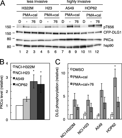
FIGURE 7.
PKCα signaling at the DLG1 scaffold is increased in highly invasive NSCLC lines relative to their less invasive counterparts. A, PKCα expression and phosphorylation of DLG1 at Thr-656 in NSCLC lines are shown. NCI-H322M, NCI-H23, A549, and HOP62 cells were transiently transfected with CFP-DLG1 and pretreated with DMSO (denoted by D) or the PKC inhibitor Gö6976 (76) for 10 min before the addition of PMA for 20 min and the phosphatase inhibitor calyculin A (cal) for the last 10 min of PMA treatment. Cells were lysed, and the lysates were probed for Thr(P)-656, DLG1, PKCα, and the loading control heat shock protein 90 (hsp90). B and C, bar graphs representing the mean ± S.E. of PKCα expression (B) and DLG1 phosphorylation at Thr-656 (C) in 8 experiments performed as described in A. Gö6976-reversible phosphorylation was calculated by subtracting the relative phosphorylation in the PMA+cal+76 condition from the relative phosphorylation in the PMA+cal condition. Significantly different from H322M cells; *, p < 0.05.

