FIGURE 2.
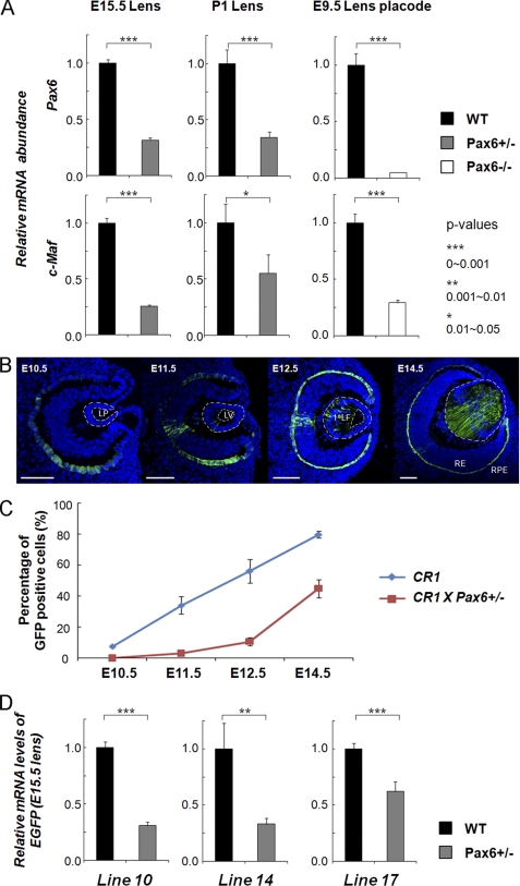
Temporal and spatial patterns of c-Maf expression in the lens are changed as a result of Pax6 haploinsufficiency. A, quantitative RT-PCR analysis of Pax6 and c-Maf expression in E15.5 and P1 wild-type Pax6+/− lenses and wild-type and E9.5 Pax6−/− lens placodes. B, EGFP expression driven by CR1/1.3kb c-Maf promoter in Pax6+/− eye. LP, lens pit; LV, lens vesicle; 1oLF, primary lens fibers; RE, retina; RPE, retinal pigmented epithelium. Scale bars = 100 μm (C and D). Blue, DAPI; green, anti-GFP. C, quantitative analysis of EGFP-positive cells generated in wild-type and Pax6+/− transgenic lenses by ImageJ. Data from three different transgenic lines were combined. D, quantitative analysis of EGFP mRNA levels of three individual transgenic lines (10, 14, and 17) in the wild type and in Pax6+/− transgenic lenses by quantitative RT-PCR.

