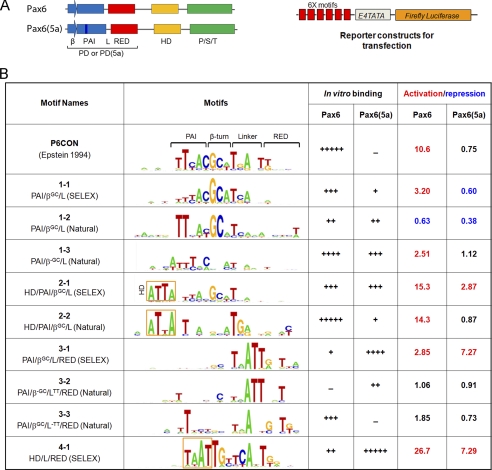
FIGURE 4.
Identification and functional characterization of novel Pax6 DNA-binding site variants. A, a schematic diagram of Pax6 and Pax6(5a) proteins and luciferase reporter constructs. β, β-turns; PAI, N-terminal subdomain of PD; L, linker region; RED, C-terminal subdomain of PD; P/S/T, transcriptional activation domain rich on serine, threonine, and proline residues. 6x motifs represent individual Pax6-binding variants. E4 TATA is a minimal promoter. B, a summary of novel variants of Pax6-binding sites. Motifs 1-1, 2-1, and 3-1 were found for Pax6. Motif 4-1 was found for Pax6(5a). Motifs 1-2, 1-3, 2-2, 3-2, and 3-3 were generated from known/validated Pax6-binding sites (supplemental Fig. S4). In vitro binding of Pax6 and Pax6(5a) to individual motifs was tested by EMSAs (supplemental Fig. S5). Cotransfection assays were performed in P19 cells, and the data are expressed as relative fold changes elicited in the presence of Pax6 or Pax6(5a) compared with the changes found with the empty vector.

