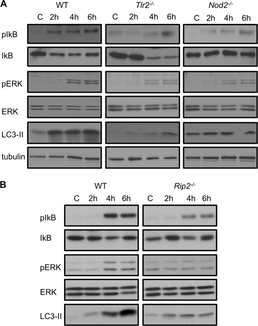
FIGURE 5.
TLR2−/− and Nod2−/− macrophages show defective NF-κB and ERK1/2 signaling upon Listeria infection. A, Western blots of pIκB, IκB, pERK, ERK, and LC3-II in WT, TLR2-, and NOD2-deficient macrophages that were uninfected (C) or infected with L. monocytogenes for different periods. Tubulin was used as a loading control. B, Western blot analyses of pIκB, IκB, pERK, ERK, and LC3-II in WT and RIP2-deficient macrophages that were uninfected (C) or infected with L. monocytogenes for different periods.

