FIGURE 7.
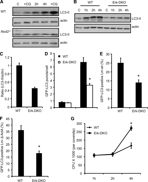
Cells deficient in ERK signaling display defective autophagy of L. monocytogenes. A, Western blot analyses of LC3-II in wild-type and Nod2−/− dendritic cells infected with L. monocytogenes. At 30 min, a set of control and L. monocytogenes-infected cells were incubated with CQ. Results are representative of three separate experiments. B, Western blot analyses of LC3-II in wild-type and Erk1−/−;Erk2fl/fl;CD11c-Cre (ERK-DKO) DCs infected with L. monocytogenes. Results are representative of three separate experiments. C, densitometry scanning of the Western blot analysis showing the ratio of LC3-II to actin in wild-type and ERK-DKO DCs infected with L. monocytogenes 4 h after infection. D, quantification of the number of GFP-LC3 puncta per cell 4 h after infection in wild-type and ERK-DKO DCs. Results show mean ± S.E. (*, p ≤ 0.05) of experiments done at least three times. E, quantification of the GFP-LC3-positive L. monocytogenes wild-type strain in cells infected as described in C. Results show mean ± S.E. (*, p ≤ 0.05) of experiments done at least three times. F, quantification of theGFP-LC3-positive Listeria mutant lacking ActA in cells infected as described in C. Results show mean ± S.E. (*, p ≤ 0.05) of experiments done at least three times. G, intracellular growth curve of L. monocytogenes ΔActA in wild-type and ERK-DKO DCs. *, p ≤ 0.05.

