FIGURE 5.
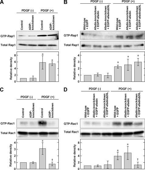
ADIP-mediated Rac activation. A, no effect of ADIP on the activation of Rap1 in NIH3T3 cells stimulated with PDGF. At 4 h after serum starvation, control or ADIP knockdown NIH3T3 cells were stimulated with or without 15 ng/ml PDGF for 1 min. Cell lysates were used for the pulldown assay and subjected to Western blotting using the anti-Rap1 pAb. B, no effect of the ADIP-afadin interaction on the activation of Rap1 in NIH3T3 cells stimulated with PDGF. The assay was performed as described in A. C, necessity of ADIP for the activation of Rac in NIH3T3 cells stimulated with PDGF. Cells were treated as described in A. Cell lysates from each NIH3T3 cell type were used for the pulldown assay and subjected to Western blotting using the anti-Rac1 mAb. D, necessity of the ADIP-afadin interaction for the activation of Rac1 in NIH3T3 cells stimulated with PDGF. The assay was performed as described in C. The bar graphs represent the relative density of GTP-bound Rap1 or GTP-bound Rac1 normalized to the total amount of Rap1 or Rac1, respectively. These values were compared with the value of the control NIH3T3 cells or wild-type NIH3T3 cells expressing EGFP without PDGF stimulation, which is expressed as 1. The error bars indicate ±S.D. *, p < 0.05 versus control without PDGF stimulation in A and C or wild type + GFP without PDGF stimulation in B and D. †, p < 0.05 versus wild type + GFP with PDGF stimulation. The results shown are representative of three independent experiments.

