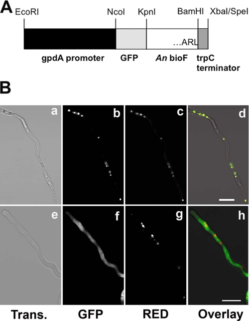
FIGURE 1.
Localization of A. nidulans BioF protein. A, the bioF open reading frame was fused to GFP, and the chimeric gene was expressed from the constitutive gpdA promoter. The BioF C-terminal amino acids are ARL. Construct GFP-BioF-FL contains full-length bioF fused to GFP (as shown in A), whereas construct GFP-BioF-TR contains bioF lacking the codons for the last three C-terminal amino acids (ARL) fused to GFP. An, A. nidulans. B, vegetative hyphae of stable transformants expressing a DsRed-PTS1 peroxisomal marker and either GFP-BioF-FL (panels a–d) or GFP-BioF-TR (panels e–h) were analyzed by confocal microscopy. Fluorescence was acquired in the green channel (GFP; panels b and f) and red channel (panels c and g). Light transmission (Trans.) images (panels a and e) and the superimposition of green and red fluorescence (panels d and h) are shown. Scale bars = 20 μm.

