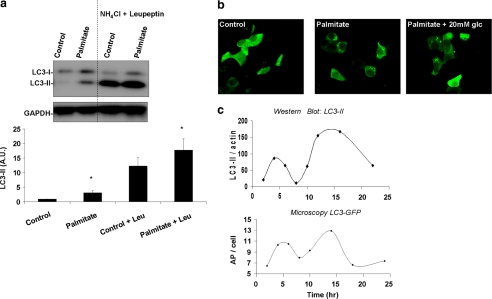
FIGURE 2.
Palmitate increases AP formation without reaching steady state. a, Western blot analysis of LC3. INS1 cells were incubated for 14 h with or without 0.4 mm palmitate, in the presence or absence of 20 mm ammonium chloride (NH4Cl) and 100 μm leupeptin (Leu) during the last 3 h of incubation. Below is the densitometry of LC3-II signal normalized against GAPDH. Note that palmitate increased LC3-II both in the absence and presence of ammonium chloride and leupeptin. *, p < 0.05 (n = 6); error bars, S.E. b, confocal images of INS1 cells expressing WIPI-GFP and incubated for 14 h in control media (containing 10 mm glucose), in media containing 0.4 mm palmitate along with 10 mm glucose or along with 20 mm glucose. c, representative curve (n = 3) of LC3-II protein levels over time as determined by densitometry of Western blot analyses. LC3-II signal was normalized against actin. Below is the average AP number per cell over time as determined from confocal images of cells expressing GFP-LC3. Between 40 and 50 cells were analyzed per condition.

