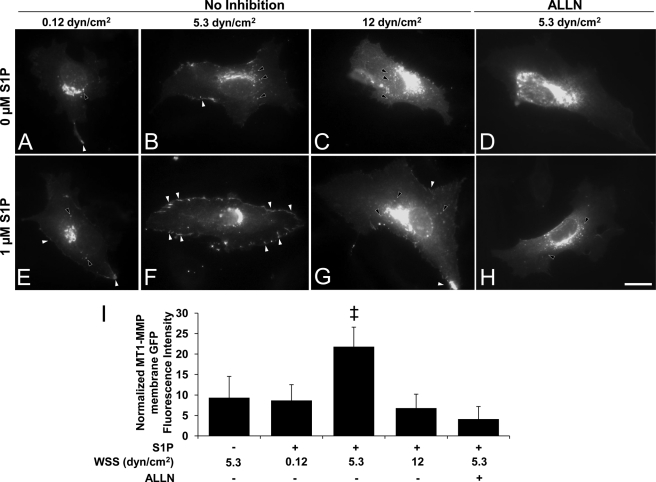
FIGURE 6.
Combined S1P and WSS treatment induced calpain-dependent membrane translocation of MT1-MMP. ECs transiently transfected with vectors expressing MT1-MMP-GFP chimeras were treated with the indicated magnitudes of WSS in the absence or presence of S1P for 2 h. Transfection efficiency was roughly 20%. In calpain inhibition experiments, cells were pretreated with the calpain inhibitor III (50 μm) for 1 h prior to S1P and WSS treatments, and the inhibitor was maintained in the perfusion medium. A–H, white arrowheads indicate MT1-MMP-GFP localization to the cell periphery; black arrowheads indicate perinuclear localization. Scale bar, 20 μm. I, MT1-MMP peripheral GFP fluorescence intensities were quantified as described under “Experimental Procedures;” ‡, p < 0.05 versus all other treatments. dyn, dynes.

