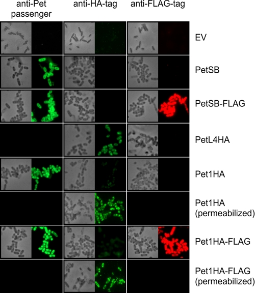
FIGURE 4.
Stalled intermediates possess a hairpin conformation that can be detected using fluorescence microscopy. TOP10 cells expressing empty vector (EV), PetSB, PetSB-FLAG, PetL4HA, Pet1HA, and Pet1HA-FLAG were collected 1 h after induction with 0.02% arabinose and subjected to indirect immunofluorescence using anti-Pet passenger antibody (left panels), anti-HA antibody (middle panels), or anti-FLAG tag antibody (right panels; pseudo-colored red) and an Alexa Fluor 488-labeled conjugate (all panels). In the case of Pet1HA and Pet1HA-FLAG, half of the samples were fixed and permeabilized prior to labeling. Corresponding fields are also shown by phase contrast microscopy. Completely black boxes indicate that staining was not assessed.

