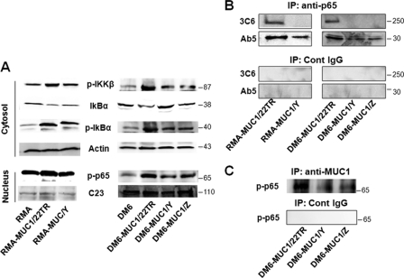
FIGURE 2.
MUC1 transfection into RMA and DM6 cell lines induces NF-κB activation and interaction with p65. A, RMA cells (left) were transfected with MUC1/22TR (RMA-MUC1/22TR) or MUC1/Y (RMA-MUC1/Y) or left untransfected (RMA) and stimulated with ConA. DM6 cells (right) were transfected with MUC1/22TR (DM6-MUC1/22TR), MUC1/Y (DM6-MUC1/Y), or MUC1/Z (DM6-MUC1/Z) or left untransfected (DM6) and stimulated with TNF-α. Expression of indicated proteins was assessed by Western blotting. Protein loading was controlled by reprobing membranes for the expression of actin and nucleolin (C23) as indicated. B, lysates from RMA-MUC1/22TR, RMA-MUC1/Y, DM6-MUC1/22TR, DM6-MUC1/Y, and DM6-MUC1/Z cells were immunoprecipitated (IP) with anti-p65 Ab or a control (Cont) IgG as described under “Experimental Procedures” and immunoblotted with anti-MUC1.CT antibody Ab5 and anti-MUC1 VNTR antibody 3C6. C, whole cell lysates from DM6-MUC1/22TR, DM6-MUC1/Y, and DM6-MUC1/Z cells were immunoprecipitated with antibody Ab5 or control IgG as described under “Experimental Procedures.” The precipitates were immunoblotted with anti-phospho-p65 antibody.

