FIGURE 1.
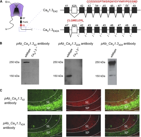
Characterization of pAb_CaV1.342 and pAb_CaV1.342A specific antibodies Western blot and immunostaining. A, left, schematic of CaV1.3 channel pore-forming α1 subunit (α1D), with hot spots for CaM/channel regulation in the carboxyl terminus (EF, EF-hand (32); pre-IQ and IQ domains (33, 34). Right, diagrammatic representation of two C-terminal splice variants of the CaV1.3 α1 subunits. Incorporation of exon 42A instead of exon 42 results in a truncated C terminus 6 amino acids after exon 41. Rabbit polyclonal antibodies were raised specifically against these two splice variants. The peptide sequences utilized are in red and italics, namely for pAb_CaV1.342, CCEDDSSPTWSRQNYSYYNRYPGSSMD, and pAb_CaV1.342A, CLQMLERL. B, Western blot of total membrane protein extracted from wild type and CaV1.3−/− knock-out mice. Left blot was probed with pAb_CaV1.342 antibody. A single band of ∼250 kDa was detected in a wild type mouse but absent in the knock-out mouse. Middle blot was probed with pAb_CaV1.342A antibody. A single band of ∼150 kDa was detected in a wild type mouse but absent in the knock-out mouse. Right blot was probed with commercial pAb_CaV1.3 antibody. C, double labeling of the CA3 region of wild type mouse dorsal hippocampus with bassoon (red) and pAb_CaV1.342 or pAb_CaV1.342A (green). Positive staining for presynaptic vesicle proteins (bassoon) is restricted to the stratum lucidum (SL), a mossy fiber recipient layer of the CA3 subfield of the hippocampus. Strong staining of pAb_CaV1.342 (top) and pAb_CaV1.342A (bottom) was observed in the stratum pyramidale (SP) of the region. No co-localization was observed between the bassoon and pAb_CaV1.342 or pAb_CaV1.342A. Scale bar, 50 μm.

