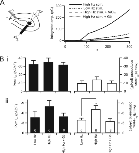
FIGURE 5.
Elevated neuronal stimulation facilitates nickel-sensitive calcium influx. A, left, a diagram illustrates the nerve stimulation and chromaffin cell recording condition. Right, evoked catecholamine release was measured by carbon fiber amperometry. Total catecholamine release was determined by integrating the amperometric record as in Figs. 1 and 4. Representative traces are plotted in B for Low Hz stimulation (stim.) and High Hz stimulation in a normal Ringer's bath solution and High Hz stimulation in the presence of Ni2+ or Gö6983-containing Ringer's bath solution. B, cells determined to be excitable by neuronal stimulation were patch-clamped in the perforated voltage clamp configuration after bipolar stimulation, and I-V protocols were conducted. Data are plotted as mean value, with error bars representing S.E. Panel i, peak inward (left axis) and the Ni2+-sensitive (sens.) inward component (right axis) currents were measured as in Fig. 3 after bipolar neuronal stimulation with Low Hz- and High Hz-stimulated firing patterns. Panel ii, mean inward current at PVm and the Ni2+-sensitive component were measured in each condition as described in Fig. 4, pooled, and plotted. * indicates p ≤ 0.05 with respect to control values determined by one-way analysis of variance. Sample size is indicated by the number at the base of each category bar. pF, picofarad; Gö, Gö6983.

