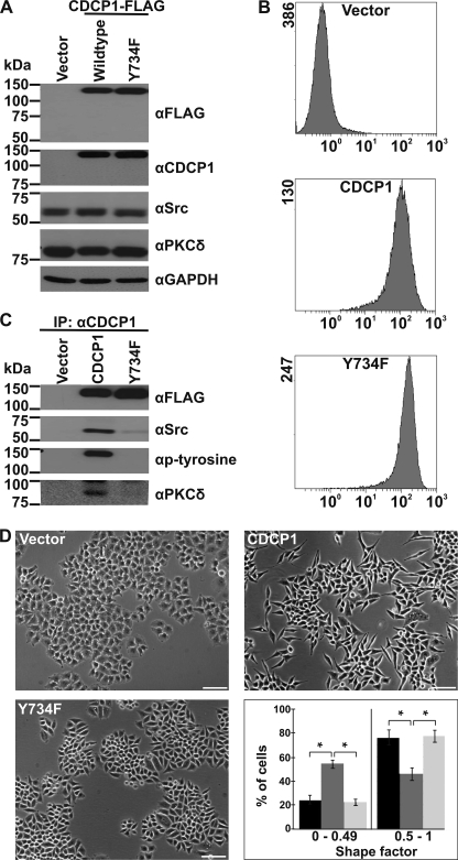
FIGURE 1.
CDCP1 expression alters HeLa cell morphology dependent on the SFK binding site. Analyses of HeLa cells stably transfected with vector or expression constructs encoding either CDCP1 or CDCP1-Y734F are shown. A, anti (α)-FLAG, -CDCP1 (10D7), -Src, - PKCδ, and -GAPDH Western blot analysis. B, flow cytometry analysis using the anti-CDCP1 monoclonal antibody 10D7. C, anti-FLAG, -Src, -phosphotyrosine, and -PKCδ Western blot analysis of proteins immunoprecipitated (IP) from HeLa vector, HeLa CDCP1, and HeLa CDCP1-Y734F cells using the monoclonal anti-CDCP1 antibody 41–2. D, transmitted light microscopy analysis. Cells were imaged using a Nikon Eclipse TE2000-U microscope (bar, 100 μm). To quantify the effect of CDCP1 expression on HeLa cell morphology, a shape factor was determined for each cell using MetaMorph software as described under “Experimental Procedures.” At least 50 cells (for each of three independent cell platings) were analyzed for each cell type, and cells were grouped into the two shape factor categories 0–0.49 and 0.5–1. These categories were graphed against the proportion of cells in each category. Black, HeLa vector cells; dark gray, HeLa CDCP1 cells; light gray, HeLa CDCP1-Y734F cells. *, p < 0.05.

