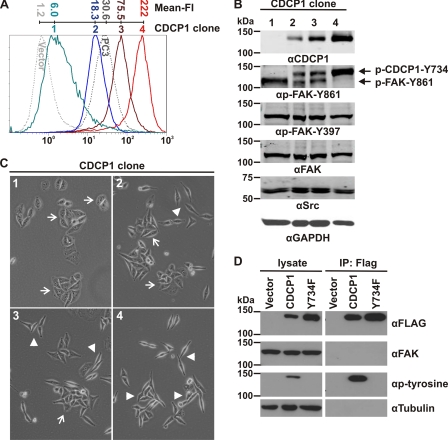
FIGURE 6.
Phosphorylation of CDCP1-Tyr-734 and FAK-Tyr-861 in HeLa CDCP1 cells is inversely related and depends on the level of expression of CDCP1, but CDCP1 and FAK do not interact. Analyses of four HeLa cell clones stably expressing different levels of CDCP1 are shown. A, anti-CDCP1 (antibody 10D7) flow cytometry analysis of four HeLa CDCP1 cell clones (numbered 1–4) that express CDCP1 at increasing levels. HeLa vector and prostate cancer PC3 cells were analyzed as negative and positive controls for CDCP1 expression (dotted lines). The mean fluorescence intensity (Mean-FI) for each cell line is shown. B, Western blot analysis of lysates from HeLa CDCP1 cell clones using anti (α)-FLAG, -p-FAK-Tyr-861 (which also recognizes p-CDCP1-Tyr-734 (20)), -p-FAK-Tyr-397, -FAK, -Src, and -GAPDH antibodies. Phosphorylated CDCP1-Tyr-734 and FAK-Tyr-861 are indicated by arrows. C, transmitted light microscopy analysis. Cells were imaged using an Olympus XX-41 microscope. Arrow, epithelial morphology; arrowhead, elongated, fibroblastic morphology. D, Western blot analysis of lysates and proteins immunoprecipitated with an anti-FLAG antibody from HeLa vector, HeLa CDCP1, and HeLa CDCP1-Y734F (clone 4) cells using antibodies against FLAG, FAK, p-tyrosine and tubulin.

