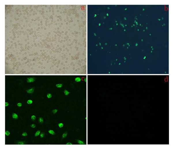
Figure 2.
Immunofluorecent staining of SSCs, detecte Oct-4 positive cells. A: After Isolating, cells observed under inverted phase contrast microscope(×100)B: Show green fluorescent cells are Oct-4 positive cells observed under Immune fluorescence microscope(×100).C: Show green fluorescent cells observed under Immune fluorescence microscope(×800).D: The negative control groups of rabbit serum replace antibody, cells observed under Immune fluorescence microscope(×100).

