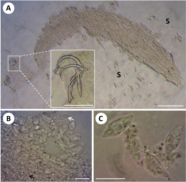
Figure 1.
Histozoic myxosporean, Myxidium incomptavermi n. sp., infecting the gill monogenean Diplectanocotyla gracilis. A) Flattened monogenean showing no internal organ definition, with myxospores (S) released from the body cavity, bar = 200 μm. Inset) fine detail of the haptoral sclerites, bar = 50 μm. B) spores released from ruptured parenchymal tissues (white arrow) with numerous spores packed inside the worm (black arrow), bar = 10 μm. C) Higher magnification of myxospores revealing a typical Myxidium morphology, bar = 10 μm. A further figure plate of air-dried spores is available from the additional file 2 S2.

