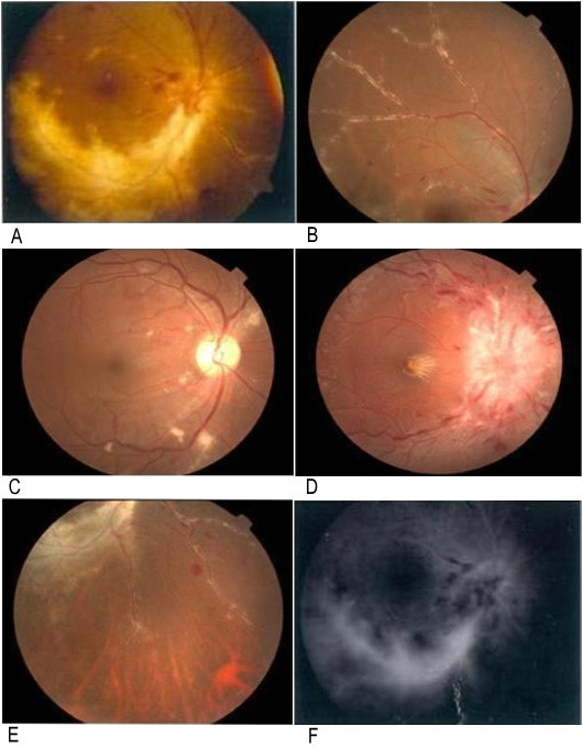
Figure 1.

Typical retinal appearance of the patients with CMV retinitis. A yellow-white retinal necrosis with granulo-margin. B retinal frosted branch angiitis. C white cotton-wool patches. D optic disc edema. E sclerosis and occlusion of retinal vessels. F fluorescence leakage by fluorescent fundus angiography.
