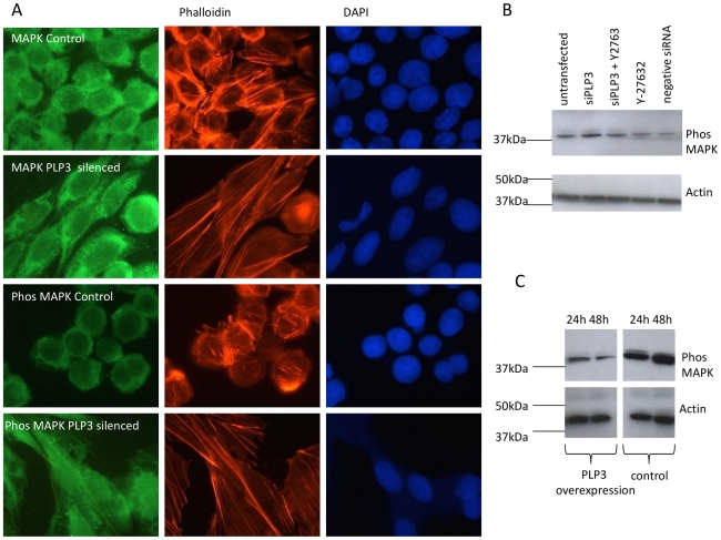
Figure 5. siRNA silencing of PhLP3 promotes MAPK phosphorylation upstream of RhoA activation and PhLP3 overexpression decreases levels of phosphorylated MAPK in suspension CHO LB01 cells.
(A) PhLP3 silenced LB01 cells were fixed and co-stained with either anti-MAPK (second panel, green) and rhodamine phalloidin (for filamentous actin, red) or anti-phosphorylated MAPK (fourth panel, green) and rhodamine phalloidin. DAPI was used to detect DNA. Untransfected cells were used as controls (first and third panel). (B) An increase in phosphorylated MAPK was detected by immunoblotting using an anti-MAPK kinase antibody in cells transfected only with PhLP3 siRNA (lane 2) compared to cells that had been transfected with PhLP3 siRNA in the presence of the ROCK inhibitor Y2763 (lane 3). Cells treated with either Y2763 only, cells transfected with a ‘scrambled’ negative control siRNA and untransfected cells did not show elevated phosphorylated MAPK levels. (C) CHO cells were transiently transfected with PhLP3 for either 24 h or 48 h and levels of phosphorylated MAPK were detected by immunoblotting with an anti-MAPK kinase antibody. Expression levels of phosphorylated MAPK were found to decrease from 24 h to 48 h (compare lanes 1 to 2) and to be lower than in untransfected cells (lanes 3 and 4).

