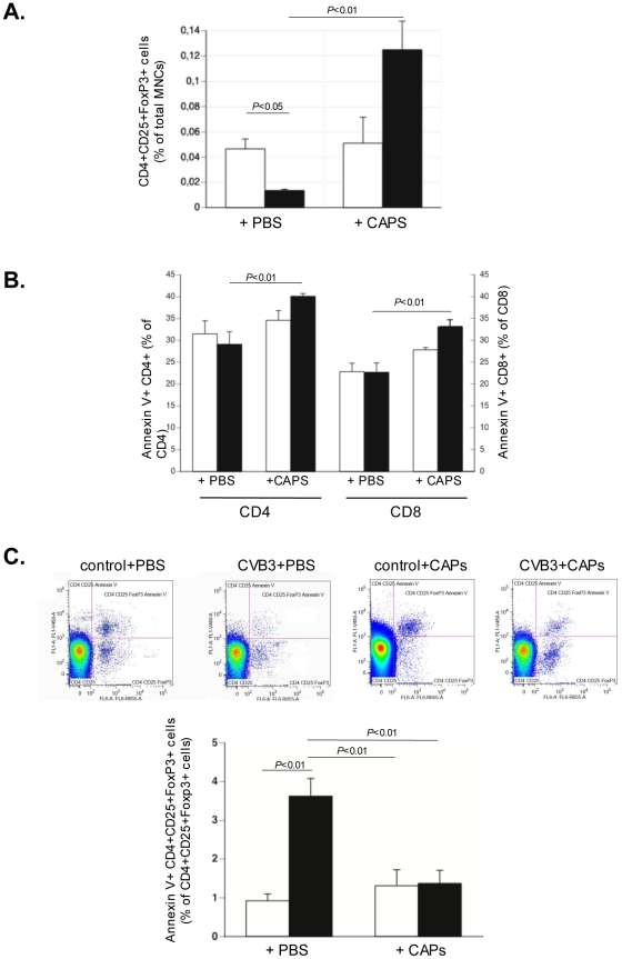
Figure 7. Cardiac adherent proliferating cells increase T regulatory cells, increase apoptotic CD4 and CD8 T cells, and decrease apoptotic T regulatory cells in Coxsackievirus B3-infected mice.
Bar graphs represent the mean ± SEM of A. T regulatory (CD4CD25FoxP3+) cells depicted as the % of MNCs, in the spleen of control mice (open bars) and CVB3-infected mice (closed bars) injected with PBS or CAPs, as indicated, with n = 4/group. B. apoptotic (Annexin V+) CD4+ and CD8 T+ cells, depicted as % of CD4+ or CD8+ T cells, respectively, in the spleen of control mice (open bars) and CVB3-infected mice (closed bars) injected with PBS or CAPs, as indicated, with n = 4/group. C. Panel demonstrates representative dot plots of CD4CD25FoxP3+ AnnexinV+ cells from preselected CD4+CD25+cells. Bar graphs representing apoptotic (Annexin V+) T regulatory (CD4CD25FoxP3+) cells depicted as the % of CD4CD25FoxP3+ cells, in the spleen of control mice (open bars) and CVB3-infected mice (closed bars) injected with PBS or CAPs, as indicated, with n = 4/group.

