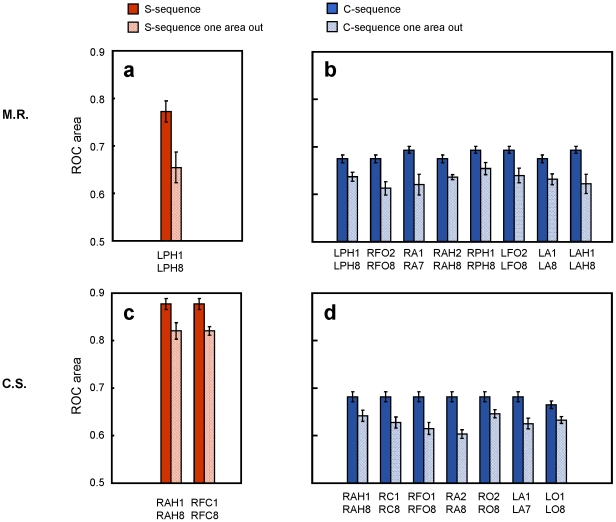
Figure 4. Localization results.
ROC areas for M.R. (a) and C.S. (c), comparing classification performance when taking into account all implanted areas versus after dropping in turn one area. Red bars refer to the S-sequence, blue bars to the C-sequence (b and d). Significant differences between keeping all the contacts and dropping in turn couples of electrodes were observed for the S-sequence only for the string including the hippocampus and right frontal caudate contact for C.S. By contrast for the C-sequence, a significant drop in the classification accuracy was evident for many areas. Importantly single-trial classification remains always lower in the C-sequence than in the S-sequence. Abbreviations: LA = left amygdala; RA = right amygdala; LAH = left anterior hippocampus; LPH = left posterior hippocampus; RAH = right anterior hippocampus; RPH = right posterior hippocampus; LFO = left fronto-orbital area; RFO = right fronto-orbital area; LO = left occipital; RO = right occipital; RFC = right frontal-caudate. Standard errors are shown.

