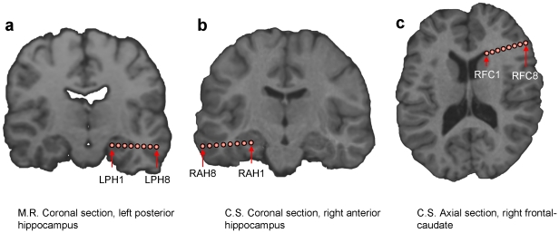
Figure 5. Visualization of the areas most contributing to the classification.
(a) Left posterior hippocampus for patient M.R. (b) Right anterior hippocampus and (c) right frontal-caudate region for patient C.S. Electrodes localized by the CT scan were coregistered to the MRI T1-weighted brain volumes.

