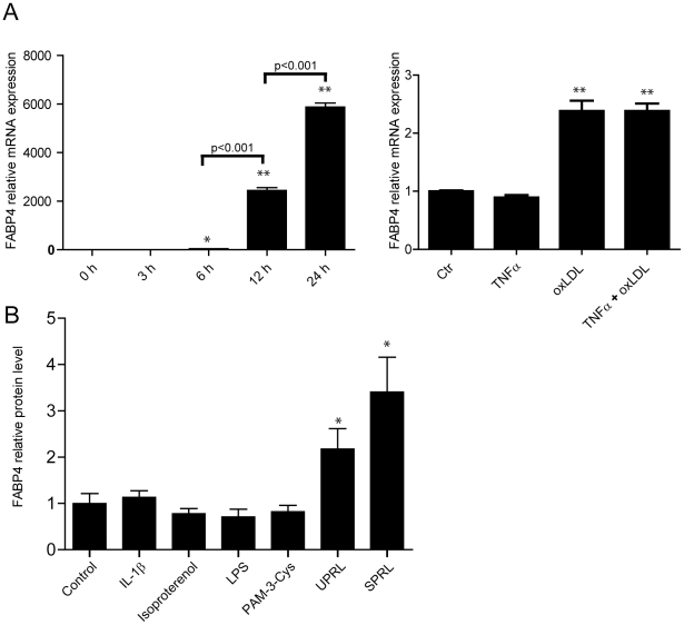
Figure 4. Oxidized LDL and platelets enhances FABP4 in macrophages.
Panel A shows mRNA expression of FABP4 in THP-1 cells during PMA differentiation (left) and in THP-1 macrophages stimulated with oxidized LDL (20 µg/ml), TNFα (5 ng/ml) or both for 18 hours. mRNA levels were quantified with the use of real-time RT-PCR. The expression of β-actin was used as endogenous control. Panel B shows the release of FABP4 protein into the cell medium, as determined by enzyme immunoassay, in THP-1 monocytes that had been pre-incubated with rhTNFα (5 ng/ml) for 96 hours before being incubated with LPS (5 ng/ml), a TLR2 agonist (Pam3Cys, 1 µg/ml), isoproterenol (20 µM), rh-IL-1β (1 ng/ml) and platelet releasate from un-stimulated (UPRL) and thrombin-activated platelets (SPRL) for additional 20 hours. Data are mean ± SEM relative to values in un-stimulated cells (control). *p<0.05 and **p<0.001 versus control (or 0 hours [h] in panel A, left).

