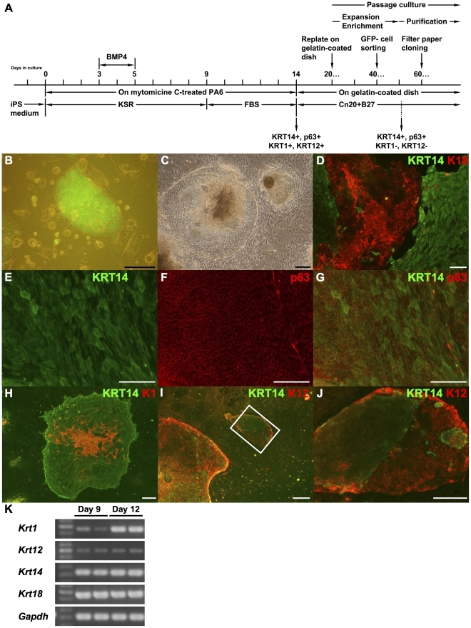
Figure 1. Epidermal and corneal epithelial cells are induced from mouse iPS cells by SDIA method.
A schematic diagram of the culture method is represented in (A). (B) Nanog-iPS cell line 38C2. Undifferentiated state was monitored by the expression of GFP. (C) Mouse iPS cells formed epithelial cell colonies on MMC-treated MPA6 cells by SDIA method with use of BMP-4 and FBS. Cell culture schedule is represented in panel K. (D) After culture days 14, the epithelial cell colonies induced by SDIA method were positive for KRT18 and KRT14. (E, F, and G) In KRT14-positive colonies, cells formed multilayer and the expression of p63 was observed especially at a high level in basal layer. The image in (G) represents a merged image of (E) and (F). Cells expressing KRT1 (H) and KRT12 (I and J) were found in KRT14-positive colonies. An image at high magnification of H is shown in I. (K) Expression of these cytokeratins were also confirmed by RT-PCR. Scale bars in B-H, 200 µM; I, 100 µM; J, 400 µM. KRT14 and p63, both stratified squamous epithelium markers; KRT18, a marker for non squamous epithelia and early surface ectoderm; KRT1, as an epidermal marker; and KRT12, as a corneal epithelial marker.

