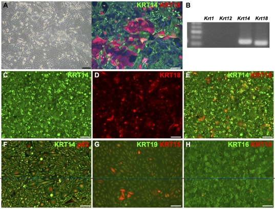
Figure 3. Characterization of mouse iPS cell-derived epithelial clone 1204SE1.
Phase contrast image (A left) and immunofluorescent image of mouse iPS cell-derived epithelial cells before cloning (A right). (B) RT-PCR analysis of the Krt1, Krt12, Krt14 and Krt18 in the epithelial cells before cloning revealed the expression of Krt14 and Krt18. (C–H) Immunohistochemical analysis of cloned iPS cell-derived epithelial cells, 1204SE1. Merged image of (C) KRT14, and (D) KRT18 is represented in (E). After cloning, all cells were positive for KRT14 and p63 (F) but negative for the epidermal marker KRT10 (I). Some cells were KRT18-positive as well. Clusters of KRT15-positive cells, a marker for basal cells of stratified epithelia, were found in 2D-cultures but cells were negative for the non-cornified squamous epithelial cell marker KRT19 (G). Cells were positive for KRT16, a marker for hyper proliferative epithelial cells (H). Scale bars, 100 µm in panel A left and C-H; 50 µm in panel A right.

