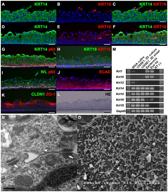
Figure 4. Characterization of cultivated epithelial sheets developed from clone 1204SE1.
Stratified epithelial cell sheets were developed from clone 1204SE1. An H&E-stained image of the five- to six-layered 3D sheet is shown in (L). The expression of cytokeratins and other epithelial markers in the 3D-cultured cell sheet were examined by immunohistochemical (A-K) and RT-PCR (M) analysis. Images are shown with the basal side down. Merged images of (A) KRT14 and (B) KRT18, and (D) KRT14 and (E) KRT10 were represented in (C) and (F), respectively. Cells were KRT14-positive throughout the epithelial sheet. Remaining KRT18-positive cells were found in the superficial layer and the expression of epidermal keratinocyte markers KRT1/KRT10 was slightly up-regulated in the stratified sheet (E and M). High expression of p63 was found in basal layers of the stratified epithelial sheet and the expression decreased in suprabasal layers (H). KRT15, which is found in basal layer of stratified epithelium, was also found in the most basal layer of the cultivated sheet (G). The cells were positive for KRT19 but negative for KRT12 (G and M). The terminally-differentiated marker Involucrin (IVL) was positive (I) and high levels of E-cadherin (ECAD) expression were found throughout the sheet (J). Tight-junction marker ZO-1 was found in the superficial layer, however, Claudin1 (CLDN1) was not observed (K). Formation of tight-junctions (arrow head in N), and abundant desmosomes (arrows in N) were observed by TEM. SEM (O) revealed formation of microvilli on the surface of epithelial sheet. Scale bars, 50 µm in A-L, 0.2 µm in N, 2 µm in O.

