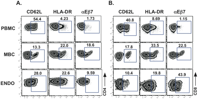
Figure 3. A representative example of surface antigen expression.

Cell surface expression of CD62L, HLA-DR and αEβ7 on A. CD3+CD4+ and B. CD3+CD8+ T cells from PBMC, MBC and endometrial tissue (ENDO) is shown. Percent of positive cells for a given marker are indicated above the gate.
