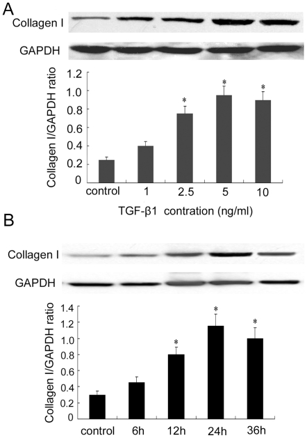
Figure 3. TGF-β1 induced collagen type I expression in a concentration- and time-dependent manner in ADPKD cyst-lining epithelial cells.
(A) TGF-β1 treatment (1–10 ng/mL, 24 h). (B) TGF-β1 treatment (5 ng/mL, 6–36 h). Top panels were representative Western blot images. Bottom panels were the summary data from three independent experiments. *P<0.05 vs. control.

