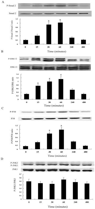
Figure 5. Time course of Smad2 (A), ERK1/2 (B), p38MAPK (C) and JNK (D) activation by TGF-β1 in ADPKD cyst-lining epithelial cells.
Cells were treated with TGF-β1 (5 ng/ml) for the indicated time periods. Kinase activation was determined by western blot analysis using phosphospecific antibodies. As controls, the protein levels of Smad2, ERK1/2, p38MAPK and JNK were determined using corresponding non-phosphorylated form antibodies. Results of densitometric analysis, expressed as a ratio between phospho- and non-phospho-antibody, from three independent experiments were shown. *P<0.05 vs. control.

