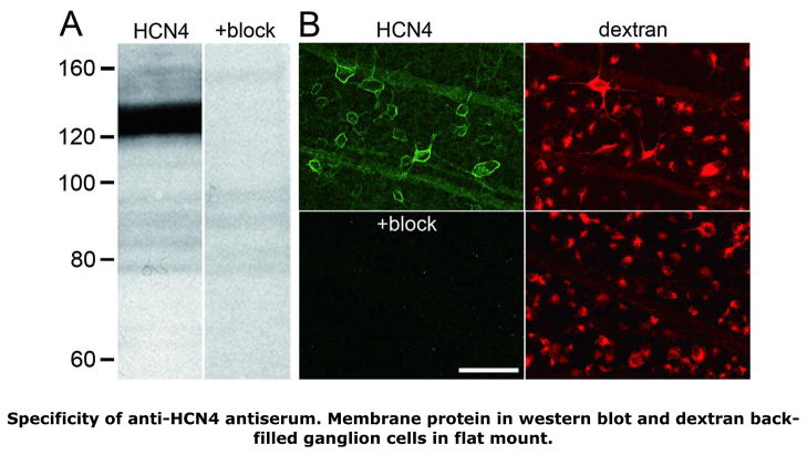
FIGURE 1.
Specificity of anti-HCN4 antiserum binding. A: Western blots of retinal membrane fraction proteins, identically prepared and formatted, except that the lane labeled “HCN4” was probed with anti-HCN4 antibody, while the lane labeled “+block” was incubated in anti-HCN4 antibody pre-incubated with control antigen. Tick marks show distance migrated in neighboring lanes by protein standards of the molecular weights indicated (in kD). The anti-HCN4 antiserum bound specifically to a band at 123 kD. B: Ganglion cell layer somata and optic fiber layer axon fascicles in two flat-mounted retinas, processed and imaged identically, except that one retina (“HCN4”) was incubated in anti-HCN4 antibody, while the other (“+block”) was incubated in anti-HCN4 antibody pre-incubated with control antigen. The ganglion cells in each retina were filled by fluorophore-coupled dextran introduced into the optic nerve, so that HCN4-like immunoreactivity (i.e., Alexa Fluor 488 fluorescence) could be imaged while focusing on the ganglion cell somata. Comparison of the “HCN4” and “+block” fields shows that control antigen blocked anti-HCN4 antibody binding. Comparison of the “HCN4” and corresponding dextran fields shows that HCN4-like immunoreactivity circumscribed ganglion cell somata and tracked axon fascicles extending across the field (between the upper left and lower right). Each image is a single confocal optical section collected with a 40x oil immersion objective. Scale bar is 50 μm and applies to all panels.

