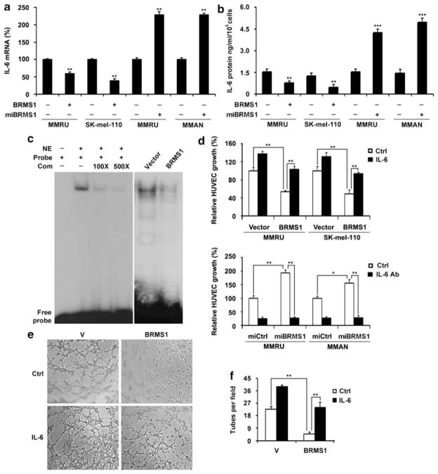
Figure 5.
BRMS1 expression in melanoma cells inhibited IL-6 expression and suppressed NF-κB activity. (a, b) BRMS1 suppresses IL-6 expression. MMRU and SK-mel-110 cells were transiently transfected with vector or Flag-BRMS1 plasmid for 48 h. In a parallel experiment, MMRU and MMAN cells were transfected with vector control or miBRMS1 plasmid for 72 h. IL-6 mRNA in the cells and secreted protein level in conditioned medium were determined with qRT–PCR (a) and ELISA (b). Data were presented as means±s.d. from three independent experiments. (c) BRMS1 overexpression suppressed the binding activity of NF-κB p65 subunit to IL-6 promoter. Competitive sequence inhibited the binding reaction in a dosage-dependent manner. (d) Addition of IL-6 recombinant protein (0.8 ng/ml) to the conditioned medium rescued BRMS1-suppressed endothelial cell growth, whereas application of IL-6 antibody (320 ng/ml) to neutralize the IL-6 in conditioned medium abrogated BRMS1 knockdown-enhanced endothelial cell growth (n=3/group). (e) Addition of IL-6 to conditioned medium rescued the tube formation inhibited by BRMS1 overexpression. (f) The numbers of tubes formed per field were counted for BRMS1-overexpressing and control groups in five random fields. *P<0.05, **P<0.01, ***P<0.001, Student’s t-test. Com, competitive sequence; NE, nuclear extract.

