
Figure 2. The normal retinal vasculature is not effected by the VE-cadherin antagonist.
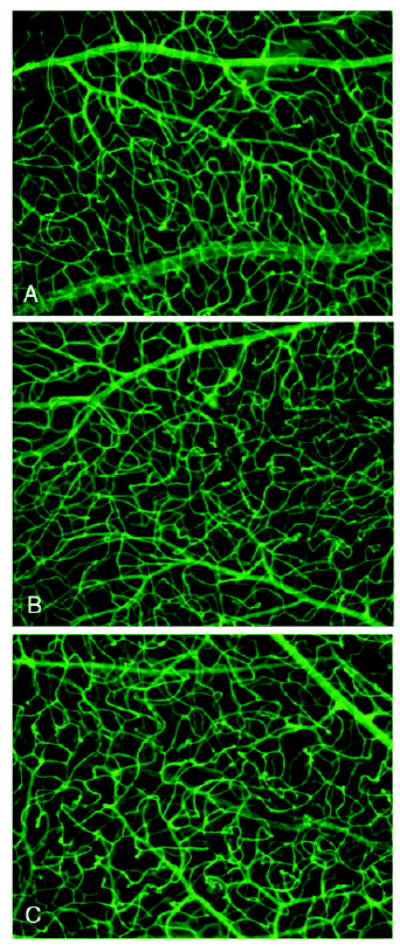
Representative images of vessels from d17 retinal whole mounts stained with FITC-GSA lectin. The vascular morphology and density of vessels within the retinal tissue is similar from animals treated with the VE-cadherin antagonist (A), the control peptide (B) or vehicle only (C).