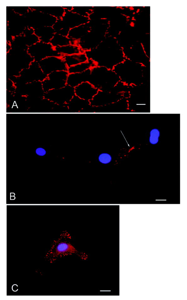
Figure 6. The VE-cadherin antagonist induces β-catenin reorganization in cultured endothelial cells.

Confluent or subconfluent bovine retinal endothelial cells were stained for β-catenin. β-catenin is present in a continuous distribution along intercellular junctions in a confluent monolayer of cells (A). Isolated cells show no β-catenin staining in the cytoplasm or at the cell membrane except where cell-cell contact is occurring (B arrow). Isolated cells incubated with the VE-cadherin antagonist demonstrate β-catenin staining both in the cytoplasm as well as at discrete locations along the cell membrane (C). Bar=20um.
