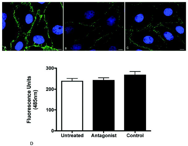
Figure 7. The VE-cadherin antagonist does not disrupt existing endothelial cell junctions.

Representative images of confluent bovine retinal endothelial cell monolayers stained for VE-cadherin in untreated (A), control peptide-treated (B) or antagonist-treated cells (C). The antagonist had no effect on the structural integrity of the monolayer as demonstrated by continuous VE-cadherin staining along the cell borders. The function of the cell junctions was assessed by measuring the permeability of the monolayer using FITC-dextran (D). The permeability of the antagonist treated cells was not significantly different from untreated cells or cells treated with control peptide. Values are the mean +/- SEM from N = 4 wells for each treatment. Bar = 10μm
