
Fig. 9.
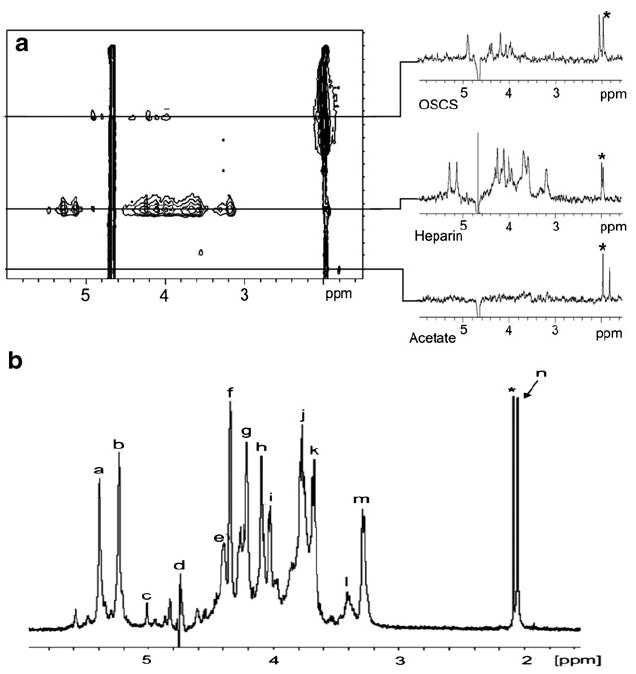
a On-flow 1H NMR-detected WAX chromatogram of heparin and OSCS. The slices taken through the chromatographic peaks show the 1H NMR spectra of OSCS, heparin, and acetate, which was present as an unexpected impurity. The asterisk represents residual acetonitrile as a column contaminant. b Stop-flow 1H NMR spectrum of heparin. The spectrum was acquired from a single 25 μL injection of a sample containing 40 mg/mL of heparin. Signals: a, H-1 of GlcNS; b, H-1 of Ido2S; c, H-1 of IdoA; d, HDO; e, H-6 of GlcNS6S; f, H-2 of IdoA2S; g, H-3 of IdoA2S; h, H-4 of IdoA2S; i, H-5 of GlcNS6S; j, H-4 of GlcNS6S; k, H-3 of GlcNS; l, H-2 of GlcA; m, H-2 of GlcNS6S; n, N-acetyl; asterisk, residual acetonitrile. Adapted from [138] with permission